Tracking worm movements using Prior Scientific’s OptiScan Motorized Stage System
Posted in Aug 25th 2020
Dr. Zhao-Wen Wang of the C. elegans Neurobiology Lab at the University of Connecticut uses the OptiScan Motorized Stage System for upright microscopes combined with his Track-A-Worm software to create an automated single-worm tracker designed for extracting and quantifying detailed bending and locomotory behavior of C. elegans.
OptiScan Motorized Stage System: Affordable solution for routine microscope applications
The OptiScan motorized stage systems provides an affordable option for users seeking an automated microscopy solution. Routine microscopy applications can be performed with much greater accuracy and speed without sacrificing either affordability and reliability. In the case of Dr. Wang and his application requirements, the OptiScan system was a perfect fit.
When Dr. Zhao-Wen Wang of the C. elegans Neurobiology Lab at the University of Connecticut needed a dependable and affordable solution for automating his research – the OptiScan system proved to be the ideal solution.
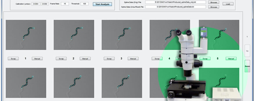
Utilizing the OptiScan controller (ES9XY), stage (ES111), and universal specimen holder (H473), Dr. Wang is able to keep a crawling worm centered in the camera imaging field. Once the worm is centered, Dr. Wang activates his Track-A-Worm software and compares positions of the worm over successive images, and uses this information to correct the stage position at 1-sec intervals. All stage movements are automatically recorded in a stage file, which is used in combination with worm image files in subsequent analyzes. Stage positions are corrected between intervals of image acquisitions. Thus there are neither blurred images nor discontinuities in the reconstructed worm path due to stage movements. The OptiScan controller, motorized stage and focus mechanism allows great flexibility and creates a powerful system tailored for Dr. Wang’s application.
The C. elegans Neurobiology Lab at the University of Connecticut focuses on worms also known as “C. elegans”, which are a common organism used in the scientific community since it is a basic organism that shares many of the essential biological characteristics that are central problems of human biology. The worms have have a nervous system with a brain that exhibits behavior and is even capable of simple learning. It produces sperm and eggs, mates and reproduces. After reproduction it gradually ages, loses strength and finally dies. C. elegans exhibits these characteristics, yet is only 1mm long and is usually grown on petri plates seeded with bacteria. All of its cells of its transparent body are visible with a microscope, and its average life span is only 2-3 weeks making the C. elegans an ideal specimen for researchers as its complex yet easy to study!
OptiScan Motorized Stage System: User Benefits
The OptiScan provides an affordable option for users seeking an automated microscopy solution. Fitting onto a wide variety of commonly used upright microscopes, the OptiScan stage provides repeatability for many routine microscopy applications. The stages can be fitted with a variety of sample holders depending on the samples being imaged. The OptiScan is flexible and its components can be configured to precisely match the users requirements and budget.
Summary:
The OptiScan system offers unsurpassed affordablity, versatility and reliability and has proven to be an ideal platform for Dr. Wang and his experiments.
For More Information:
https://health.uconn.edu/worm-lab/wp-content/uploads/sites/136/2017/09/User-manual-v1.pdf