Life Science Microscopy with the PureFocus850
Posted in PureFocus 850 Jan 10th 2024
In this article, we discuss compatibility of the PureFocus850 with transmitted brightfield, transmitted darkfield and fluorescence microscopy, as well as the advantages of having a hardware autofocus installed on a microscope. Other important microscopy techniques for life science will be discussed in the next article, so keep an eye out or contact us if you would like to know more about a different technique.
What are the advantages of hardware autofocus in microscopy?
Hardware autofocus systems are extremely useful in life science microscopy. They can be used to increase throughput of imaging or ensure samples remain in focus for the duration of a long timelapse experiment.
They use reflection of a laser signal from the sample to determine its position in space, and then automatically adjust the microscope focusing (Z) axis to bring the sample back into focus.
This gives them three key advantages over camera-based autofocus solutions:
- They do not rely on contrast within the sample, meaning that fluorescent or unstained samples can be kept in focus more easily.
- Focus capture is extremely rapid and is done in real time, whereas camera-based solutions require multiple seconds of scanning via the microscopes Z axis.
- Hardware autofocus solutions track the sample surface, so there is no requirement to pre-define a focus map at different points in the sample.
Prior’s PureFocus850 is an extremely versatile hardware autofocus with numerous features to allow life science microscopists to take advantage of these benefits, across a broad spectrum of microscopy techniques and sample types. Critically for life science microscopy, the PureFocus850 can track the surface of coverslips while maintaining focus on the sample below.
How does hardware autofocus adapt to various microscopy techniques?
Brightfield Microscopy
Transmitted (diascopic) brightfield microscopy of thin sections is arguably the most routine microscopy technique. Simply, white light passes through the sample, and variance in light absorption and scattering across the different features produces an image. ‘Brightfield’ refers to the near-white background contrasted against the darker features of the sample itself.
The PureFocus850 features an offset mechanic, which allows focus on the sample to be maintained by tracking the controlled surface of a coverslip – an essential feature for imaging even the most standard slides. Life science samples are weakly reflective, and as such the coverslip is detected by the PureFocus850. The offset mechanic allows the Purefocus850 to focus on the coverslip while the microscope eyepieces or camera are focused on the sample.
However, some applications, such as blood cell analysis, require high magnification oil immersion objectives to resolve sample features in sufficient detail. This prevents the outer surface of the coverslip from being used by the PureFocus850 because immersion oil and glass have very similar refractive indices. The offset, however, can be adjusted to allow the inner surface of the coverslip to be detected instead.
Brightfield microscopy typically involves the use of colour camera with high detection efficiency in the visible light spectrum: the PureFocus850 laser uses an infrared wavelength to avoid any potential for the laser signal appearing on the image of the sample.
Darkfield Microscopy
Transmitted darkfield microscopy has many applications within life science. Most commonly it is used to observe tissue sections or cells which have low inherent contrast which make brightfield microscopy an unsuitable observation technique.
Darkfield microscopy relies on the reflection or refraction of light passing through the sample, which allows it to be collected by the objective. Any light that is not affected by the sample is not collected. This causes the background to be dark (hence the name darkfield!) and samples to appear bright. This in turn allows intracellular detail to be observed without staining the sample, or small particles or contaminants in the sample to be more easily detected.
As the PureFocus850 does not rely on contrast to obtain focus, the often-sparse signal produced by darkfield microscopy does not affect performance. Darkfield is also prone to allowing objects outside of the sample plane to appear brightly in the image and camera-based autofocus techniques can cause focus erroneously on these objects. Because the PureFocus850 relies only on the coverslip for focusing, it avoids these errors entirely.
Fluorescence Microscopy
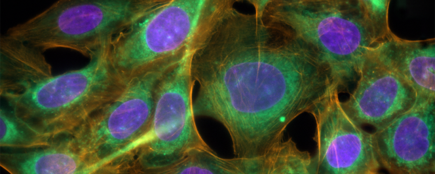
While darkfield microscopy can provide contrast in transparent samples, they do not highlight specific features and are limited with how small the highlighted features can be. Fluorescence microscopy, either via transformation of live cells or organisms or labelling of sections, in contrast allows localisation and identification of specific organelles other subcellular features.
Fluorescence is a phenomenon where an object, typically a molecular label conjugated to a protein in the context of microscopy, absorbs light at one wavelength and emits it at a longer wavelength. This differentiation between absorbed and emitted signal allows light exclusively emitted by the fluorescent molecular label to be detected, if the microscope is fitted with optics to only allow light of this wavelength to reach the detector. The overall result is that these specific features are now strongly highlighted against a black background.
Multiple features with different fluorescent labels can be imaged in the same sample. Motorized devices such as filter wheels and turrets can move the correct excitation filters (to select the absorption wavelength) and emission filters (to select the emission wavelength) into the microscope light path, and images taken in sequence. A composite image of the different labels can then be generated.
Applications for fluorescence microscopy are wide-ranging, from relatively simple widefield techniques such as fluorescence in situ hybridisation (FISH) or high throughput screening, to more complex high end microscopy methods such as spinning disk confocal microscopy and total internal reflection microscopy (TIRF).
Typically, fluorescent labels require excitation light between the near ultraviolet and far-red wavelengths. As a result, any hardware autofocus must not interfere with any wavelengths being used to produce the image. The PureFocus850 uses an 850nm laser, which sits beyond the emission wavelength of Cy7, a common fluorescent label that emits close to the near infrared spectrum. Additionally, the PureFocus850 transmits near-UV and visible light, enabling it to be positioned between the fluorescence optical components and the objective. This is important as it allows complete flexibility in the choice of optical components when configuring a fluorescence microscope.
How can Purefocus850 be adapted to other high-end microscopy techniques?
Although we discussed many of the most popular life-science microscopy techniques, the list is not exhaustive. The PureFocus850 can be modified for use with several other high-end microscopy techniques – such as multiphoton microscopy – which use middle UV or infrared light and may require specific optical materials or coatings.
Interested in PureFocus850 for your application?
If you have a specific application not mentioned here, please contact us to discuss your requirements. In our next blog post, we will discuss compatibility of the PureFocus850 with phase contrast techniques, differential interference contrast and polarisation microscopy.