Raman and Infrared Microscopy for Chemical Mapping
Automating Raman and infrared microscopy for high-throughput research
Raman and infrared microscopy are both used to study the chemical composition and molecular structure of samples. They each provide different types of information about molecular bonds and are often used together as complementary techniques to give users a more comprehensive analysis of materials.
Raman microscopy relies on Raman scattering to generate images. While most photons scatter randomly (Rayleigh or elastic scattering), a few scatter with a shift in energy (inelastically), corresponding to molecular vibrations. Unlike most microscopy techniques, Raman microscopy does not generate images of the sample’s physical structure; instead, it provides information about the sample’s molecular composition. Typical applications include:
- Life science: Visualize cell components without staining.
- Materials science: Map stress in semiconductors or composite materials by revealing the molecular structure.
- Drug discovery: Create false-color chemical maps to reveal the chemical composition of tablets.
Infrared microscopy is a catch-all term for microscopy using infrared wavelengths of light. This might be transmitted infrared imaging of semiconductors, single photon fluorescence imaging using infrared-absorbing and emitting fluorophores, or multi-photon imaging of tissues by multiple harmonic-generation.
The range of applications is extremely broad, with some typical examples being:
- Life science: Mapping fluorescently labelled proteins deep in tissue samples or organoids.
- Materials science: Visualize additive distribution in plastics or inspect both sides of semiconductor wafers.
- Label-free imaging: Using the inherent properties of connective or muscle tissue to image via second-harmonic generation (SHG) or third-harmonic generation (THG).
Challenges in Raman microscopy
Focus Drift During Mapping
This can occur due to large-area scans or uneven samples. The solution is to use a laser autofocus system to maintain sharp focus throughout mapping.
Prior Scientific Solution:
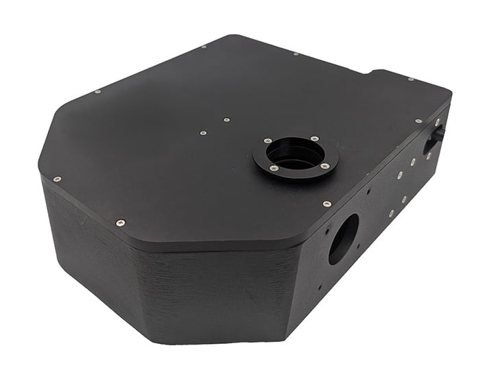
PureFocus690 Laser Autofocus System
The PureFocus690 is optimized for infrared imaging applications and offers excellent transmission of light across a broad spectral range. It supports wavelengths up to 1100 nm in the near-infrared region, while also accommodating all commonly used Raman microscopy excitation wavelengths, such as 532 nm, 633 nm, and 785 nm—making it highly versatile for both IR and Raman microscopy.
Long Acquisition Times
Chemical mapping creates an image where each pixel represents the chemical composition at that point. High-resolution chemical maps require thousands of spectra: a 100 x 100 pixel map contains 10,000 spectra, each of which may take up to a second or more to acquire if the signal is weak. Manually positioning the samples is also slow, subjective, and error-prone.
Automating the process frees operators to focus on reviewing and analyzing the data.
Prior Scientific Solution:
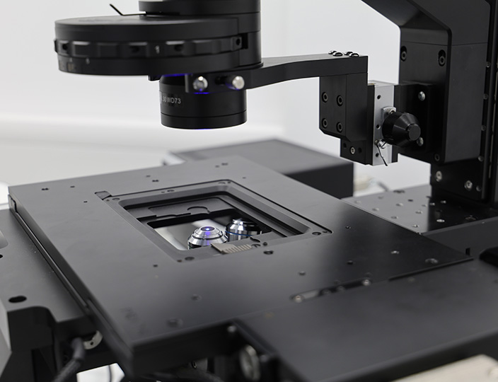
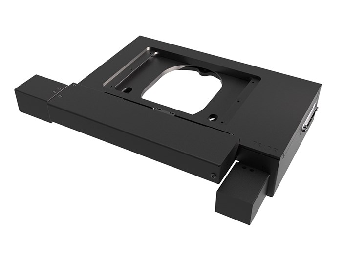
ProScan Motorized XY stage with Intelligent Scanning Technology
A highly stable stage that can maintain its position reliably is important for any application with long acquisition times.
Prior’s H117 High Performance Stage for Inverted Microscopy or H101N1F Flat Top High-Resolution Stage for Upright Microscopy both offer sub-micron resolution. The combination of stepper motors and ball screws provides sufficient friction to keep the stages static during acquisition without compromising motion.
Prior’s ProScan stages feature Intelligent Scanning Technology (IST), which ensures movement accuracy without the need for encoders, and prevents oscillations at the end of moves.
Queensgate’s NanoScan SP Z Series of nanopositioning Z-axis piezo stages are highly stable during long acquisition times due to their capacitive sensor technology. The piezo stages offer market-leading velocity, step-settle time, and sub-nanometer resolution.
Challenges in IR microscopy
Thickness Variations
While IR is very good at penetrating thick samples, imaging within organoids or tissues with the right level of speed and precision is challenging.
Prior Scientific Solution:
Queensgate Z-axis piezo nanopositioning stages
Queensgate’s piezo nanopositioning stages offer sub-micron step-scanning on the z-axis with long travel ranges up to 800 µm to enable imaging within thick samples. They are highly stable and, with <10 ms step-settle times, facilitate the capture and reconstruction of 3D images, and are compatible with a wide range of inverted and upright microscopes.
Life science samples for IR imaging, such as organoids in media or whole animals, often require the sample to be completely stable. This means that the objective, rather than the sample, should be moved for refocusing. NanoScan OP series of nanopositioning objective scanners control the movements of the objectives. They offer exceptional repeatability and resolution and have a load capacity up to 1000 g, enabling them to support the large and heavy objectives typically used for multiphoton imaging.
NanoScan SP Z Series of nanopositioning Z-axis piezo stages are slim and compatible with a wide range of samples including well-plates, petri-dishes and slides. They are particularly useful when requiring high precision movement for multiple objectives but the sample is immobile, for example when imaging brain sections.
Scanning large samples
Inspection of semiconductors with IR can enable detection of various properties and defects within individual chips and can save time by imaging both sides of the wafer simultaneously. Such large samples, however, are inherently time consuming to image completely.
Prior Scientific Solution:
ProScan H112 large format stage with PureFocus690 laser autofocus system
The ProScan H112 motorized XY stage has a 300 x 300 mm travel range, which is large enough for fully mapping 12-inch silicon wafers. We offer a range of mounting accessories, including vacuum or chuck holders for wafers, breadboards, and aluminum plates.
Maintaining focus across the large wafer surface area while scanning is another challenge. This is where the PureFocus690 works with the large-format stage, keeping the sample in focus by adjusting to changes in sample height in real time. This can reduce scanning times by up to 95% by removing the need for contrast based autofocus or volumetric imaging. The PureFocus690 also has a motorized offset lens to switch the focus plane between the upper and lower surfaces of the wafer while the autofocus is active.
Sample drift during time-lapse experiments
Stability is key during the extended acquisition times involved in time-lapse experiments. No matter how stable the equipment set up is, sample drift is a real challenge, especially with live cell imaging, where changes in the sample can change the focal plane considerably.
Prior Scientific Solution:
The PureFocus690 Laser Autofocus System provides real-time focus control to automatically adjust to changes in the sample due to thermal instability. By tracking a reflective interface in the sample, such as the water/glass boundary in a culture dish, a hardware autofocus can maintain focus on even weakly contrasted samples such as transgenic zebrafish over long periods.
Experiment apparatus variation
Multiphoton systems often image slides/tissues and whole animals, using specialized objectives designed for high-precision imaging deep into samples. With large samples and additional equipment, space can be a real challenge.
Prior Scientific Solution:
Our physiology platforms feature large surfaces to mount large samples and peripheral equipment, while allowing access for micromanipulators.
The H189 and HZ106 high-precision XYZ decks offer precise movement in the XY and Z axes. With up to 50mm of motorized Z travel the same microscopy set up used to image fluorescently labelled brain sections can be transformed to enable neuroscience experiments on whole mice in moments. For more complex experiments, the platform that the sample is mounted on can be repositioned higher or lower within the deck system, making these systems extremely flexible and ideal for research.
Image Credit: Creator: Sonjajnos, Licenced under Creative Commons ShareAlike 4.0 International

Related Products
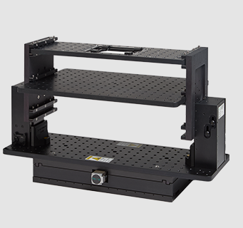
H189 High Precision XYZ Deck
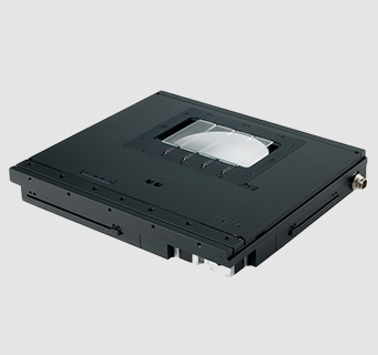
ProScan H101N1F Motorized XY Stage
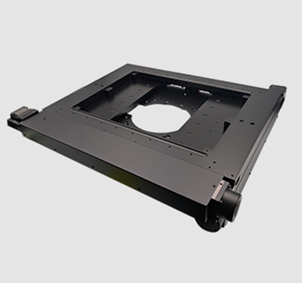
ProScan H112/2ST Large Format Motorized XY Stage
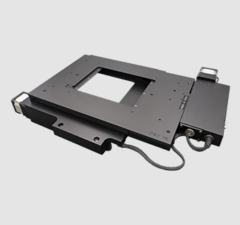
ProScan H117P1N4 Motorized XY Stage
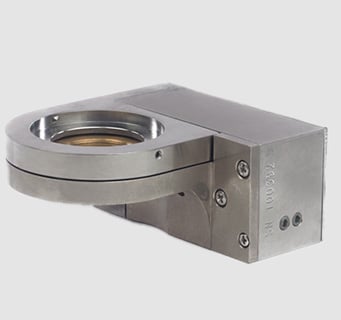
NanoScan OP400 Piezo Objective Scanner
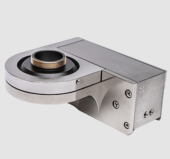
NanoScan OP800 Piezo Objective Scanner
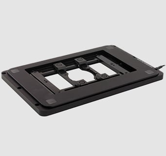
NanoScan SP Z Series Piezo Z-Axis Stages
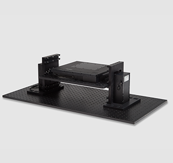
HZ106 Motorized XYZ Deck
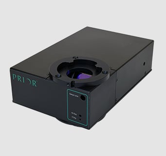
PureFocus™690 Laser Autofocus System
Articles