• Skip to primary navigation
  • Skip to main content
  • Skip to footer
Prior Scientific

Prior Scientific

Experts in Precision Positioning and Microscope Automation

  • Products
    • Automated Imaging Systems
      • OpenStand-U
      • OpenStand-M
      • OpenStand-L
      • OpenStand-V
    • Imaging Components
      • Motorized Stages
      • Focus Mechanisms
      • Laser Autofocus
      • Filter Wheels, Turrets & Shutters
      • Controllers & Joysticks
      • Robotic Sample Loaders
      • Physiology Platforms
      • Motorized Vertical Stages
    • Microscope Automation
      • Evident Olympus Microscope Automation
      • Nikon Microscope Automation
      • Leica Microscope Automation
      • Zeiss Microscope Automation
      • Automated Custom Imaging Systems
      • Other Microscope Brands
    • Nanopositioning Devices
    • Vibration Control
    • Custom Solutions & Contract Manufacturing
  • About
  • Applications
  • Blog
  • Contact Us
  • News
  • Technical Support
  • US Shop
  • 日本語
    • English
    • Deutsch
    • United States
    • 简体中文
  • Automated Imaging Systems
    • OpenStand-L
    • OpenStand-M
    • OpenStand-U
    • OpenStand-V
  • Imaging Components
    • Motorized Stages
    • Focus Mechanisms
    • Laser Autofocus
    • Filter Wheels, Turrets & Shutters
    • Controllers & Joysticks
    • Robotic Sample Loaders
    • Physiology Platforms
    • Motorized Vertical Stages
  • Microscope Automation
    • Evident Olympus Microscope Automation
    • Nikon Microscope Automation
    • Leica Microscope Automation
    • Zeiss Microscope Automation
    • Automated Custom Imaging Systems
    • Other Microscope Brands
  • Nanopositioning Devices
  • Vibration Control
  • Custom Solutions & Contract Manufacturing
Home » Applications » Pathology

Pathology Workflow Automation

Automated microscopy and advanced imaging techniques for pathology

Efficient pathology and histopathology workflows 

Microscopy is central to pathology. It helps specialists detect abnormalities, classify diseases and guide treatment decisions. Histopathology involves staining samples to improve contrast under white light – gaining clear visualization of tissue structures.  

For many pathology applications, a simple microscope is more than adequate. Small, cost-effective systems are widely used in many laboratories and bring excellent results.  

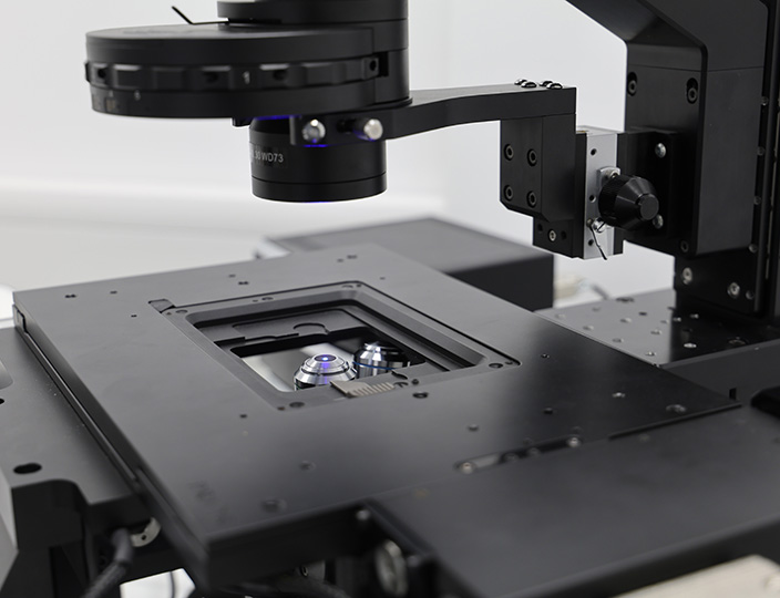
But for many workflows, automation upgrades can bring big benefits. New research- and clinical-grade microscopes are often fully or partially motorized, and existing equipment can be upgraded with motorized components for more precise imaging, operator time savings and smoother systems. 

Challenges in pathology workflows 

Manual microscopy – while accurate – can be slow and labor-intensive. Examining multiple samples under high magnification requires careful adjustments and prolonged focus – leading to fatigue and strain for operators.  

Similarly, manual imaging brings the potential for variability, making it harder to maintain consistency across large batches. It also does not allow for digitization or remote control. That’s where automation can help. 

Automating a manual microscope for pathology 

Automation adds to a manual microscope’s capabilities in many ways. For example, motorized components can help improve precision, helping to find areas of interest more easily and streamline workflows, making repetitive tasks more manageable and reducing operator fatigue.  

These upgrades are also widely compatible with existing microscopes. Laboratories can modernize without replacing entire systems. Some of the potential upgrades include: 

  • Focus control – Automatically maintains sharp images, reducing the need for manual adjustments while improving imaging accuracy. Manual focus controls are also a significant point of user fatigue.  

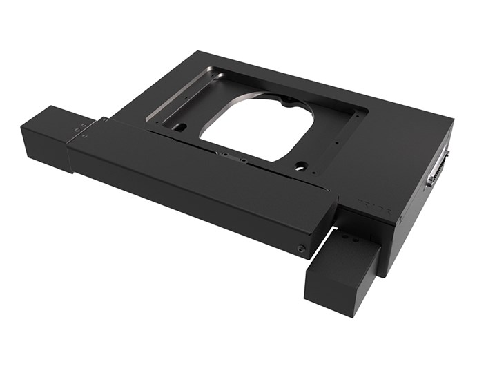
  • Motorized stage – Allows for smooth, controlled, accurate sample movement, making it easier to scan and locate areas of interest with high precision. 

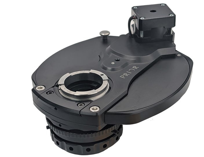
 Fluorescence accessories – adding filter wheels, alongside a multi-bandpass dichroic filter cube, can enable automated fluorescence on microscopes fitted with a manual turret.  

The benefits of microscopy automation 

Protect existing investments 

Upgrading a standard microscope with automation extends its usefulness. When combined with suitable software, it also avoids the need for costly research-level replacements. Many automation components integrate with existing manual systems, making this an accessible option for laboratories looking to modernize workflows without a substantial overhaul. 

Reduced operator fatigue 

Extended periods of manual operation cause strain and discomfort. Potential upgrades include motorized joysticks that reduce repeated wrist movements, allowing operators to control focus and stage movement with minimal effort. Likewise, flat-top stages give easier access to samples, improving user comfort and making workflows more straightforward. 

Digitization of slides 

Digital pathology removes the need for physical slide storage, giving near-instant access to images for review and comparison. Prior Scientific’s stages, equipped with our Intelligent Scanning Technology (IST), deliver precise and repeatable positioning to keep each scan highly accurate. This helps ensure that digital images are perfectly stitched and easy to analyze.  

Remote access 

A fully motorized system also supports remote collaboration. Experts can review samples from different locations – far from the microscope. Remote access also assists in teaching environments, with students controlling an instrument from another location. 

Applications of automation in pathology 

Some of the applications of microscope automation in pathology include: 

  • Hematology – Blood analysis requires high magnification to identify individual cells and bacteria. As such, precision movement is essential to avoid errors when working at these levels. Motorized stages – such as the H101E1F– allow for stable and repeatable positioning and high accuracy, essential when working with high magnification objectives.  

  • Cytology – Fluorescence microscopy is used in cytology to distinguish different tissues using fluorescent markers. Laboratories with manual fluorescence microscopes can upgrade them with motorized components to improve imaging efficiency and consistency.  

  • Phase contrast and DIC – These techniques help identify key structures in samples. Combined with artificial intelligence (AI), they allow for the increased automation of analysis and less extensive sample preparation. 

  • Slide scanning – Applies automation and digitization on a larger scale, capturing entire slides as high-resolution digital images. This improves efficiency in high-throughput laboratories, simplifies sample storage and enhances data sharing.  

Improve your pathology workflow 

Pathology laboratories face increasing demands for speed, accuracy and consistency. Manual microscopy, while effective, brings many challenges in precision, efficiency, and ergonomics. Automation is a simple, cost-effective upgrade option. Using various techniques, it makes microscopy more precise, easier to use, and less strenuous for the operator.  

If you’re looking to buy a new manual microscope and add motorization – or upgrade an existing system – Prior’s pathology automation solutions are widely compatible with many leading brands, such as Nikon and Evident. 

With upgrades like motorized components, automated focus control and digital imaging, laboratories can build more reliable, intuitive, adaptable workflows. Contact our experts to learn more. 
Enquire Now
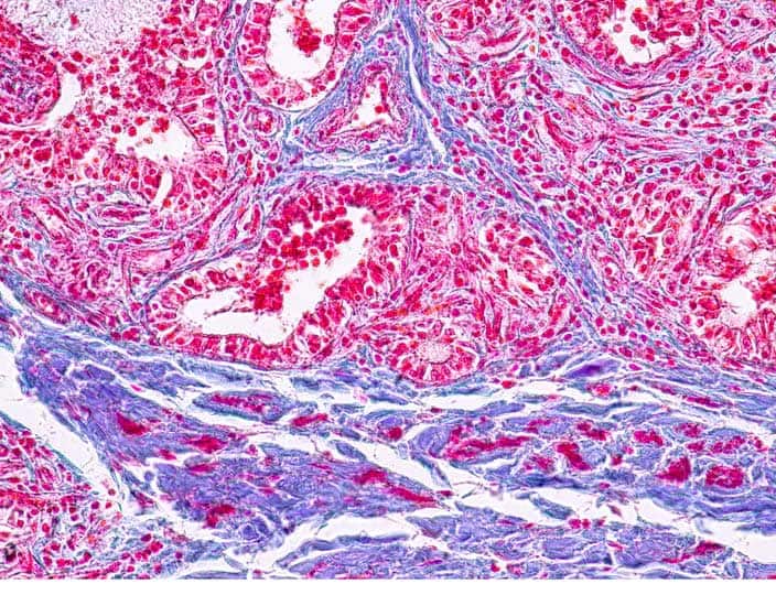
Stained pathology sample.

Related Products

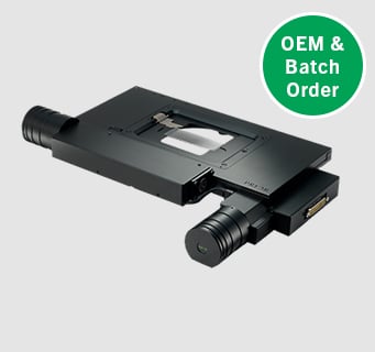
H101A stepper motor XY stage for upright microscopes with OEM/batch order flash.

ProScan H101A Motorized XY Stage

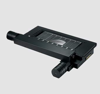
ProScan H138A motorized stage with extended lateral travel for imaging large samples or multiple slides.

ProScan H138A Motorized XY Stage

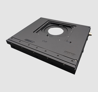
H101 Flat Top Motorized XY Stage for High Speed Upright Microscopy

ProScan H101N2F Motorized XY Stage

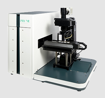
SL160 Robotic Slide Loader For High Throughput Screening.

SL160 Robotic Slide Loader For High Throughput Screening

View All Products

Articles

Close up of a motorized xy stage on an OpenStand-V inverted imaging platform. imaging platform
27/07/2026

Microscopy and Precision Positioning Glossary: Terms, Definitions and Industry Guide

↘ View Article
H100A compact precision motorized XY stage for upright microscopes
15/06/2026

H100A Compact Motorized XY Microscope Stage for Precision Microscopy: Product Manager Q&A

↘ View Article
Three quarters view of the Prior motorized filter cube turret for multi-colour fluorescence imaging.
13/05/2026

How a motorized filter cube turret expands microscope performance across advanced imaging workflows

↘ View Article
21/04/2026

HF108B Filter Wheel: High‑Speed Fluorescence Imaging for Automated Microscopy Systems

↘ View Article
View Articles

Footer

Products

  • Automated Imaging Systems
  • Imaging Components
  • Microscope Automation
  • Nanopositioning Devices

Company

  • Custom Solutions & Contract Manufacturing
  • Applications
  • Technical Support
  • Careers

Resources

  • About
  • Blog
  • News
  • Contact Us

    Sign up to stay in the know...

    ✉

    Connect with us

    Prior Scientific Instruments Ltd.
    Units 3-4 Fielding Industrial Estate
    Wilbraham Road, Fulbourn
    Cambridge, CB21 5ET, UK

    inquiries@prior.com
    +44 (0)1223 881711

    Prior Scientific Inc.
    80 Reservoir Park Drive
    Rockland, MA. 02370
    U.S.A.

    info@prior.com
    +1 781 878 8442

    Prior Scientific Instruments GmbH
    Maria-Pawlowna-Str. 4
    07743 Jena
    Germany

    jena@prior.com
    +49 (0)3641 242 010

    Prior Scientific KK
    Kayabacho 3rd Nagaoka Bldg 10F,
    2-7-10, Nihonbashi Kayabacho
    Chuo-Ku, Tokyo 103-0025, Japan

    info-japan@prior.com
    +81 (0)3 5652 8831

    Prior Scientific Instruments (Suzhou) Ltd.
    Room 118, Meilihua Hemu Park,
    No. 393 Suhong Middle Road,
    Suzhou Industrial Park,
    Suzhou, 215000, China

    info-china@prior.com
    +86 (0)512 6617 5866

    Copyright © 2026 · Prior Scientific. Terms & Conditions | Privacy Policy | Impressum

    Manage Consent
    To provide the best experiences, we use technologies like cookies to store and/or access device information. Consenting to these technologies will allow us to process data such as browsing behavior or unique IDs on this site. Not consenting or withdrawing consent, may adversely affect certain features and functions.
    Functional Always active
    The technical storage or access is strictly necessary for the legitimate purpose of enabling the use of a specific service explicitly requested by the subscriber or user, or for the sole purpose of carrying out the transmission of a communication over an electronic communications network.
    Preferences
    The technical storage or access is necessary for the legitimate purpose of storing preferences that are not requested by the subscriber or user.
    Statistics
    The technical storage or access that is used exclusively for statistical purposes. The technical storage or access that is used exclusively for anonymous statistical purposes. Without a subpoena, voluntary compliance on the part of your Internet Service Provider, or additional records from a third party, information stored or retrieved for this purpose alone cannot usually be used to identify you.
    Marketing
    The technical storage or access is required to create user profiles to send advertising, or to track the user on a website or across several websites for similar marketing purposes.
    • Manage options
    • Manage services
    • Manage {vendor_count} vendors
    • Read more about these purposes
    View preferences
    • {title}
    • {title}
    • {title}