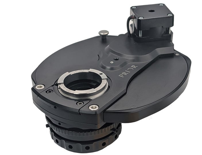
Access Confocal Imaging with Affordable Equipment
03.03.26
Researchers can view biological samples with unprecedented detail using confocal microscopy. It overcomes many limitations of fluorescence microscopy such as blurry images caused by fluorophores above and below the focal plane; inability to image thick samples; and poor contrast in highly fluorescent samples. In addition, confocal microscopy enables optical sectioning in thick samples, allowing researchers to generate sharp optical 'slices' through thick samples; three-dimensional images can be created by layering a series of these slices; and, because only the focal plane is illuminated, the risk of photobleaching is reduced.
However, a typical confocal imaging system is significantly more expensive and complex than a widefield fluorescence microscope. This can put the technique out of the reach of many researchers without the budget to invest in new equipment.
Is it possible to build your own confocal microscope?
The good news is that it’s entirely possible to upgrade existing microscopes using components from Prior Scientific and CrestOptics to create a high-end imaging system at a fraction of the cost of investing in a new, off-the-shelf confocal microscope.
Here's one we made earlier:
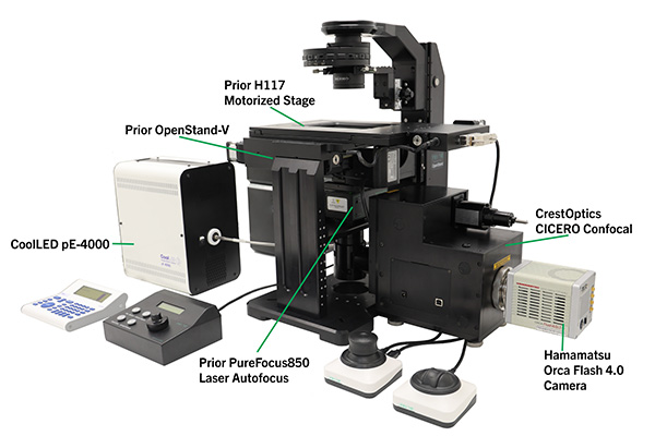
Description of the system:
Microscope:
We used the Prior OpenStand-V inverted modular imaging system as the base microscope for our confocal. Because it is modular, the Prior OpenStand offers limitless opportunities to configure and upgrade the system to meet the changing needs of users looking for a custom microscope. It is also an ideal prototyping platform for testing out and developing new imaging techniques with a view to full commercialisation.
Our example system is equipped with a motorized XY stage, laser autofocus, a motorized nosepiece, and transmitted light. It can be further enhanced with a piezo Z-stage for fine focus control, additional light paths, as well as filter wheels and turrets for other imaging modalities. It is also compatible with Nikon, Evident, Zeiss, and other types of optics, allowing users to pick the best option for their work, rather than compromising.
Motorized XY Stage
Confocal imaging generates complex datasets that are often stitched together to form three-dimensional images of large samples. Precise, repeatable automated positioning is essential to achieve high-quality results. As well as smooth motion, the stage needs to be stable – this is especially important for time-lapse imaging on live samples. The stage is available with optional linear encoders to further improve its long-range linearity: an essential feature for time-lapse imaging.
Prior’s ProScan H117 Motorized XY Stage for Inverted Microscopes features a 1 mm ballscrew and 400-step motor drive configuration for high-resolution movements together with stability. It features Prior’s Intelligent Scanning Technology to optimize stage accuracy and linearity.
As with all Prior’s motorized XY microscope stages, it comes with a full five-year warranty.
Laser Autofocus: Prior PureFocus850
Laser autofocus is essential for confocal imaging because it keeps the sample perfectly focused throughout long or demanding acquisitions, preventing image drift caused by mechanical or thermal changes in the system. We installed the PureFocus850 in our system to maintain the correct distance between the objective and reference interface in real time, ensuring consistent, high‑quality fluorescence imaging. Its 850 nm infrared laser operates outside the wavelengths typically used for confocal excitation, avoiding the risk of interference, while its built-in dichroic is specifically chosen to pass fluorescence emission wavelengths.
Confocal: Crest Optics CICERO
The CICERO from Crest Optics is a complete widefield fluorescence imaging and spinning-disk confocal unit, that is compatible with most upright or inverted microscopes. It has been used both with Prior’s OpenStand-U and OpenStand-V systems. It supports wide spectral ranges and works with either laser or LED illumination, giving users the flexibility to choose the most appropriate light source and fluorescent dyes for their application.
The CICERO’s compact size makes it a good companion for OpenStand, allowing it to take responsibility for the entire illumination and detection capabilities of the system, or be used in conjunction with other imaging modalities via the OpenStand’s epi- or diascopic illumination pathways.
Illumination: CoolLED pE-4000
In this system, we chose CoolLED’s pE-4000 as a high-power LED source for the confocal imaging, due to laser safety considerations. The pE‑4000 offers a highly adaptable illumination platform that can complement confocal workflows, particularly in systems that combine laser‑based scanning with widefield or auxiliary fluorescence modes. With its broad selection of LED wavelengths distributed across four independently controlled channels, it provides flexible excitation options for sample screening, fluorophore validation, and multimodal imaging. It has 16 discrete LED channels and built-in cleanup filters for excitation, and is an excellent option for bright, fixed samples.
Camera: Hamamatsu Orca Flash 4.0
We chose the Hamamatsu Orca Flash 4.0 Digital CMOS camera to capture the confocal images. It is highly sensitive to low light and able to detect low-signal confocal samples and has a fast frame rate – up to 100 frames per second – to support live dynamics and fast scanning.
Other options for confocal imaging
Z Drive: Piezo focus devices from Queensgate
Piezo focus devices offer millisecond settling times, enabling rapid acquisition of Z-stacks. This speed is critical for confocal volume imaging.
Queensgate nanopositioning piezo Z stages provide exceptional speed, precision, and stability for confocal microscopy, making them ideal for high‑resolution, high‑throughput imaging. Their sub‑nanometer positioning accuracy ensures crisp optical sectioning, while fast step‑and‑settle times enable rapid z‑stack acquisition without sacrificing image quality. Because Queensgate stages use closed‑loop capacitive feedback sensors, they deliver highly repeatable motion and long‑term drift stability—critical for demanding techniques such as live‑cell imaging, super‑resolution workflows, and quantitative 3D reconstruction. Their compact, low‑profile design also integrates easily into existing microscope platforms, allowing researchers to enhance imaging performance without compromising optical path geometry or sample accessibility.
The NanoScan SP Z Series of sample positioners is compatible with Prior’s motorized XY stages, including the H117, and offers 200 µm, 400 µm, 600 µm and 800 µm closed-loop ranges. Queensgate’s NanoScan OP range of piezo objective positioners is compatible with most objectives.
Laser source: 89 North LDI-7
Using a laser source for confocal imaging offers benefits that often outweigh the additional safety considerations. Spinning-disk confocal imaging is a lossy technique, and the high power of the illumination lasers mitigates this problem. By their nature, lasers need very little wavelength cleanup to excite specific fluorescence labels.
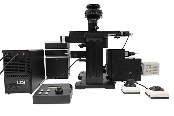
In this alternative system, we used 89 North’s LDI-7 Series solid-state laser illuminator. It offers up to 1000 mW of output power per laser to provide a brighter signal for dim samples and faster overall imaging times.
89 North’s LDI-7 is commonly used alongside the CICERO in applications such as live cell imaging, where the speed of spinning-disk confocal imaging is required. For system integrators, the LDI-7's interlock system could be coupled to Prior’s ProScan III controller to create a laser-safe complete instrument.
Talk to us about your application.
This automated confocal imaging system is just one example of how Prior’s microscope automation components can transform existing equipment into a high-end imaging system to help labs maximize their budgets. Contact us to learn more.
More Blogs
Microscopy and Precision Positioning Glossary: Terms, Definitions and Industry Guide
Learn More
H100A Compact Motorized XY Microscope Stage for Precision Microscopy: Product Manager Q&A
Learn More
How a motorized filter cube turret expands microscope performance across advanced imaging workflows
Learn More